Conventional treatment of pressure sores can be unsatisfactory – it has been shown that more than 35% of grade I pressure sores fail to heal and their condition worsens [Arashi et al, 2010]. Could vibration therapy be an alternative treatment for pressure sores or an adjunct to conventional therapy? Research offers such hope. According to a study by Arashi et al [2010], 15-minute local vibrotherapy treatments, applied three times a day for several days, significantly supported standard treatment of grade I pressure sores.
Pressure sores mainly affect people with mobility problems – the severely ill or elderly – and just how big a problem they are is made clear by data provided by Guest et al [2018], who estimate that the average cost of treating pressure sores in the UK alone is approaching £1.5 million per day! Moreover, these costs will increase as the population ages.
Conventional treatment of pressure sores is not satisfactory
In situations where pressure sores are at risk, it is important to provide appropriate care to prevent their formation or progression. In the treatment of stage I pressure ulcers, the main care is to reduce the pressure forces on the body to ensure sufficient blood supply and to prevent damage to the skin surface. This is done by changing the position and applying dressings with suitable protective preparations. In many cases, however, the deterioration of pressure sores cannot be stopped. Mainly due to the lack of appropriate ways to increase tissue microcirculation [Arashi et al., 2010].
Vibration can have beneficial effects on local microcirculation
There are physiotherapeutic techniques, such as the automated administration of vibration with appropriate parameters (vibrotherapy), that improve circulation (including microcirculation), both in healthy people and those with circulatory problems.
Ren et al [2019] (Beihang University, China) showed that in diabetic and control healthy patients, a 5-minute administration of mechanical vibration with a frequency of 50 hertz (Hz) and an amplitude of 2 millimetres (mm) significantly increased blood flow through the skin of the soles of the feet during and after the intervention.
Around 16 years earlier, also Zhang et al [2003], researchers from the Department of Orthopaedics at Sahlgrenska University Hospital (Sweden), observed a beneficial effect of vibration with variable frequency parameters (5-2000 Hz) and acceleration (16-46 m/s2) on blood flow, which they increased by 20 percent. They studied blood flow in the vessels of the tibialis anterior muscle when vibration was applied to the foot.
Vibration can promote wound healing
In the journal Acute Care, our editorial colleague published a case study of a patient suffering from venous ulcers in 2018. After 2-week vibrotherapy using the Vitberg + Rehabilitation Massage Apparatus, there was a 98% reduction in ulcer area and an average wound reduction of 1.4 cm2 per day [Pasterczyk, 2018]. Details can be found in the article: Vibrotherapy in the treatment of ulcers and chronic wounds. A case study.
Others have also drawn attention to the regenerative properties of mechanical vibration in the context of wound healing or even fracture healing (for more on the latter, see the article ‘Vibration models macrophage function, enhances early inflammatory response and promotes healing of osteoporotic fractures’ on our Specialist Portal), with macrophages – immune-competent cells – playing an extremely important role in the mechanisms underlying these processes.
Researchers from the United States (Pongkitwitoon et al [2016]) showed in vitro studies on cell cultures that mechanical vibration regulates macrophage proliferation and phenotype and healing processes. As they noted – macrophages responding correctly to biochemical signalling pathways are essential for tissue healing, as the nature (phenotype) of macrophages can be twofold: more conducive to either repair or pro-inflammatory processes, depending on environmental signals. Thus, therapies that modulate the phenotype and function of macrophages may influence healing processes.
Pongkitwitoon et al [2016] subjected macrophages to vibration stimulation to test whether macrophages could receive such signals at all and whether they would affect changes in the expression of genes encoding proteins involved in tissue repair. They found that vibration significantly increased the number of macrophages after 3 days of stimulation. Vibrations of 100 Hz and 0.15 g gave the greatest response. Also, the expression of genes encoding pro-growth factors VEGF and TGF-β increased after vibration. In addition, vibration (100 Hz and 0.15 g) significantly reduced the levels of the pro-inflammatory cytokines IL-6, IFN-γ and TNF-α (by about 10% on day 1 and by about 40-50% on day 3).
Thus, these results demonstrated the sensitivity of macrophages to mechanical vibration signals with specific parameters and may suggest that vibration may promote tissue repair processes by reducing inflammation and promoting a healing-promoting macrophage phenotype [Pongkitwitoon et al., 2016].
Vibrotherapy can support the treatment of pressure sores
We can now return to the findings of Arashi et al [2010], with whom we began this study. Following reports of the beneficial effects of vibration on circulation, including improved microcirculation in tissues, Arashi et al [2010] attempted to develop a new methodology for the treatment of pressure sores using therapeutic vibration.
In their desire to develop an effective clinical tool, they focused on the safety and clinical efficacy of local vibrotherapy, i.e. applied to selected parts of the body. In doing so, they hypothesised that local vibrotherapy would increase local venous flow and accelerate healing of stage I pressure sores.
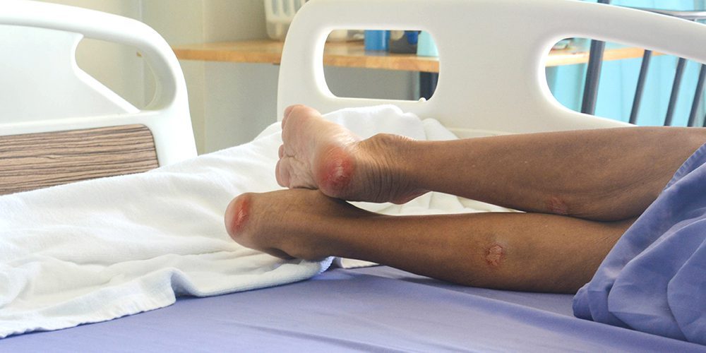
They studied 31 long-term care patients (age: over 65 years) with stage I pressure sores. The vibration group of patients (vibration group, 16 patients, n = 16) was treated from June to September 2006 and the control group (no vibration, n = 15) from October 2006 to January 2007. All patients received the same standard nursing care. The only difference between the treatment of the vibration group and the control group was that the vibration group was given additional vibration and the control group was not. Vibration was given for 15 minutes, 3 times a day, up to a maximum of 7 days or until healing of stage I sores, if it occurred earlier. The number of healed ulcers was analysed, as well as the rate of healing.
Use of vibration in the study by Arashi et al [2010].
The researchers used a RelaWave vibrator (Matsuda Micronics Corp, Japan). The size of the vibrator was 61.6 x 18.2 x 11.4 cm (length x width x height). A vibration frequency of 47 Hz and a horizontal acceleration of 1.78 m/s2 was used.
Patients who were lying down were treated. If the decubitus ulcer was on the lower limb, the vibrator was placed between the bed mattress and the pillow adjacent to the limb, while in other cases, the vibrator was placed under the mattress – between it and the bed frame. The vibrator, however, was always located under the decubitus ulcer in question. Vibrotherapy was not carried out until 2 hours after meals, and the interval between vibration sessions was at least 2 hours.
Results obtained by Arashi et al [2010].
- Vibrotherapy increased the healing rate of decubitus ulcers: wound area and the intensity of redness decreased significantly faster compared to standard treatment.
- After vibrotherapy, 40.0% (eight) of the bed sores healed, while 9.5% (two) healed after standard therapy.
- No physical discomfort caused by vibration was noted during the study.
Summary
Arashi et al [2010] conclude that local vibrotherapy can facilitate the treatment of stage I pressure sores.
Together with earlier and later reports on the improvement of blood circulation by vibrotherapy (including in diabetic patients), its promotion of wound healing, as well as fractures, we would like to draw the attention, not only of patients, but especially of rehabilitation therapists and physiotherapists, to the clinical potential of applying vibration to the area of pressure sores.
Further and urgent research should serve to develop an optimal treatment method for this disorder affecting many people in long-term care units, but also those immobilised in home beds or wheelchairs, who are waiting for help right now.
Complied from:
Arashi M, Sugama J, Sanada H i wsp., 2010. Vibration therapy accelerates healing of Stage I pressure ulcers in older adult patients. Adv Skin Wound Care 23(7):321-327. https://pubmed.ncbi.nlm.nih.gov/20562541/
Chow SK, Chim YN, Wang J, Zhang N, Wong RM, Tang N, Leung KS, Cheung WH, 2019. Vibration treatment modulates macrophage polarisation and enhances early inflammatory response in oestrogen-deficient osteoporotic-fracture healing. Eur Cell Mater 7(38):228-245. https://www.ecmjournal.org/papers/vol038/pdf/v038a16.pdf
Dube A, Sidambe V, Verdon A, Phillips E, Jones S, Lintern M, Radford M, 2021. Risk factors associated with heel pressure ulcer development in adult population: A systematic literature review. J Tissue Viability 28:S0965-206X(21)00115-7. https://www.sciencedirect.com/science/article/pii/S0965206X21001157?via%3Dihub
Guest JF, Fuller GW, Vowden P, Vowden KR, 2018. Cohort study evaluating pressure ulcer management in clinical practice in the UK following initial presentation in the community: costs and outcomes. BMJ Open 25;8(7):e021769. https://pubmed.ncbi.nlm.nih.gov/30049697/
Pasterczyk A, 2017. Wibroterapia, jako forma terapii wspomagającej w leczeniu owrzodzeń i ran przewlekłych – opis przypadku. Ostry Dyżur 10(2):64-69. http://ostry-dyzur.net/wp-content/uploads/2018/08/Str.64-69.pdf
Pongkitwitoon S, Weinheimer-Haus EM, Koh TJ, Judex S, 2016. Low-intensity vibrations accelerate proliferation and alter macrophage phenotype in vitro. J Biomech 21;49(5):793-796. https://pubmed.ncbi.nlm.nih.gov/26897645/
Ren W, Pu F, Luan H i wsp., 2019. Effects of Local Vibration With Different Intermittent Durations on Skin Blood Flow Responses in Diabetic People. Front Bioeng Biotechnol 7:310. https://www.frontiersin.org/articles/10.3389/fbioe.2019.00310/full
Zhang Q, Ericson K, Styf J, 2003. Blood flow in the tibialis anterior muscle by photoplethysmography during foot-transmitted vibration. Eur J Appl Physiol 90(5-6):464-469. https://link.springer.com/article/10.1007/s00421-003-0904-5